


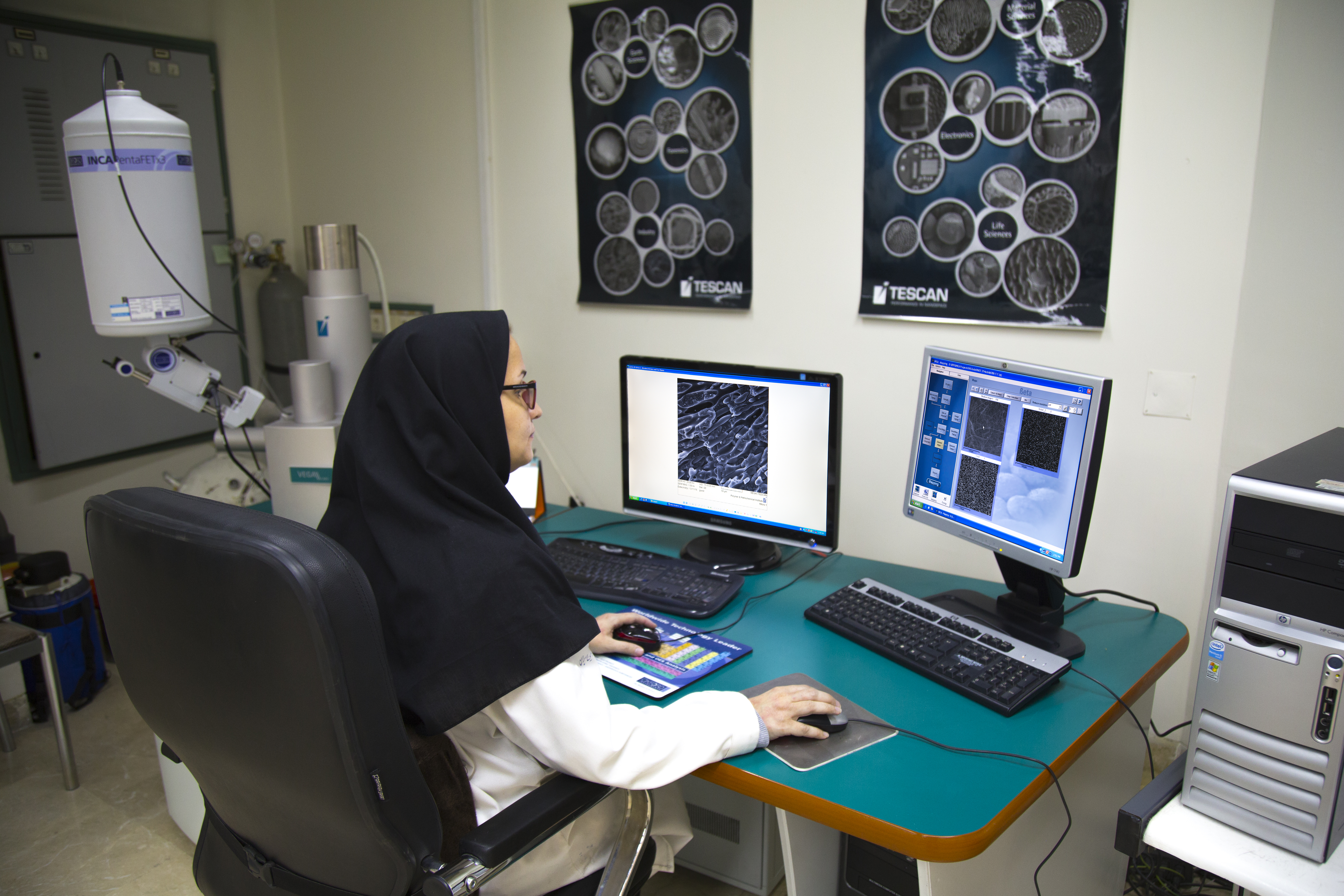
Optical and Scanning Electron Microscopy Laboratory Scaning electron microscope The SEM is a type of electron microscope that images a sample by scanning it with a high-energy beam of electrons in a raster scan pattern. The electrons interact with the atoms and produce signals that contain information about the sample's surface topography, composition, and other properties such as electrical conductivity. The types of signals produced by the SEM include secondary electrons, back-scattered electrons (BSE) and characteristic X-rays originated from the atoms at or near the surface of the sample. In the most common secondary electron imaging, the SEM can produce very high-resolution images of a sample surface, revealing details less than a few nanometers in size. Due to the very narrow electron beam, SEM micrographs have a large depth of field yielding a characteristic three-dimensional appearance useful for understanding the surface structure of a sample.
The optical microscope, often referred to as the "light microscope", is a type of microscope which uses visible light and a system of lenses to magnify images of small samples. The image from an optical microscope can be captured by normal light-sensitive cameras to generate a micrograph/or by a charge-coupled device (CCD) cameras to allow the capture of digital images. The following techniques are performed in this lab: Stereo microscope, a low powered microscope which provides a stereoscopic view of the sample, Inverted microscope, for studying samples from below; useful for cell cultures in liquid, Polarizing microscope, whose design usually includes a polarizing filter, rotating stage and gypsum plate to facilitate the study of minerals or other crystalline materials whose optical properties can vary with orientation, hot stage facility to study the phase transitions upon sample heating and cooling and Vickers hardness measuring device. The laboratory is equipped with the following devices:
|
طراحی سایت توسط پورتال aryanic